Research

Overview

The Quantitative biology of cell growth team is engaged in research on several fundamental questions related to the control of proliferation and aging in the model organism Saccharomyces cerevisiae. Our main research axes aim at i) unravelling the mechanisms that drive the entry into replicative senescence in budding yeast, ii) apply quantitative analyses to decipher the complexity of the physiological response to oxidative stress and starvation in yeast.

The lab develops microfluidics devices for non-destructive single-cell imaging in controlled conditions that, combined with molecular genetic methods, allow to monitor the dynamics of these processes at the single cell scale.

Dynamics and mechanism of entry into replicative senescence in yeast

Budding yeast cells undergo a limited number of divisions before entering senescence and eventually dying, a phenomenon known as replicative aging. Over the last twenty years, classical genetic analyses have identified genes and pathways that regulate this process, yet a detailed understanding of the cascade of events driving the entry into senescence is largely missing. In this context, we and others have pioneered the development of microfluidics-based techniques to trap individual mother cells and to monitor their successive divisions from birth to death under the microscope. Using this methodology, we have shown that cells undergo an abrupt transition (referred to as the Senescence Entry Point, or SEP) to a slow division regime that precedes cell death.


Following up on these observations, we have recently shown that the SEP is mechanistically associated with the accumulation of extrachromosomal rDNA circles (ERCs), a long described hallmark of aging in yeast. In addition, we have shown ERCs fuels an excessive transcription of ribosomal RNAs that accumulate in the nucleus, thereby compromising nuclear homeostasis.

Based on this, we are now trying 1) to better understand how the stability of the rDNA is impaired and drives the excision of ERCs from the rDNA locus and 2) to unravel the mechanism driving nuclear homeostasis control in aging mother cells with large pools of ERCs.

Quantitative Signaling during the response to oxidative stress

Unicellular organisms have evolved exquisite defense mechanisms to buffer against variations in internal physiological parameters or to counteract unpredictable environmental changes. The response of oxidative stress (e.g. hydrogen peroxide) is a fundamental and highly conserved regulatory system that allows the cell to ensure a precise redox balance, which is essential for proper physiological function. Yet, although the molecular players involved in the response are well characterized at the biochemical level, how these components integrate into a functional homeostatic system remains quite obscure.

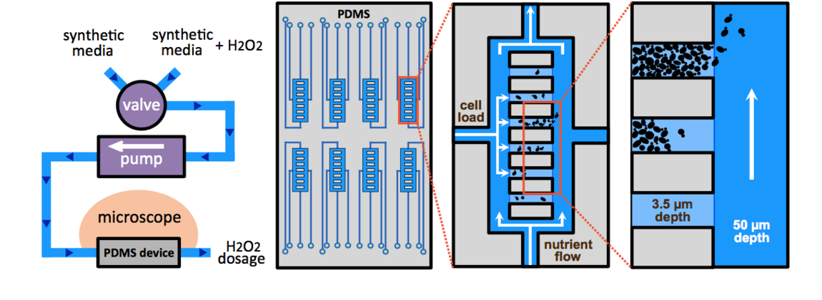
In this context, we have developed a microfluidic platform to monitor the physiological response of individual cells to various temporal stress patterns (i.e. steps, ramps, etc.) and we have deciphered the functional properties of the redox homeostatic system. We have shown that cell survival is highly stress-rate dependent: all cells consistently die when abruptly exposed to a given dose of hydrogen peroxide, yet can survive 10-fold higher doses when progressively exposed to the same stressor. This observation revealed that adaptation is limited by the response time of the homeostatic machinery, and unraveled an unprecedented ‘trainability’ of the cells to stress. We demonstrated that this particular feature, as well as the long described acquisition of tolerance, are mediated by key H2O2 scavenging enzymes called peroxiredoxins.

Following on this functional characterization of the response to oxidative stress, we are now trying to understand how cell reallocate their resources from cell growth to H2O2 scavenging when facing an oxidative stress challenge, knowing that some metabolites (e.g. NADPH) are involved in both anabolic processes and redox control.

Development of microfluidic devices for single cell dynamics assays

Our research greatly rely on the development of custom-made microfluidic devices to monitor the growth of individual cells over multi-generation timescales. We are currently developing microfluidic devices to improve the acquisition of data during the entry into replicative senescence and to combine live cell imaging data with complementary biochemical analyses of the response to oxidative stress. We have set up a clean room that allows us to perform the standard SU8-based photolithography processes in order to generate microfluidic prototypes.


Dynamics of response to starvation

Coming soon...